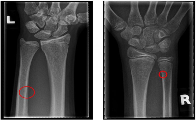
The paper proposes a segmentation and classification technique for fracture detection in X-ray images. This novel rotation-invariant method introduces the concept of local entropy for de-noising and removing tissue from the analysed X-ray images, followed by an improved procedure for image segmentation and the detection of regions of interest. The proposed local Shannon entropy was calculated for each image pixel using a sliding 2D window. An initial image segmentation was performed on the entropy representation of the original image. Next, a graph theory-based technique was implemented for the purpose of removing false bone contours and improving the edge detection of long bones. Finally, the paper introduces a classification and localisation procedure for fracture detection by tracking the difference between the extracted contour and the estimation of an ideal healthy one. The proposed hybrid method excels at detecting small fractures (which are hard to detect visually by a radiologist) in the ulna and radius bones—common injuries in children. Therefore, it is imperative that a radiologist inspecting the X-ray image receives a warning from the computerised X-ray analysis system, in order to prevent false-negative diagnoses. The proposed method was applied to a data-set containing 860 X-ray images of child radius and ulna bones (642 fracture-free images and 218 images containing fractures). The obtained results showed the efficiency and robustness of the proposed approach, in terms of segmentation quality and classification accuracy and precision (up to 91.16% and 86.22%, respectively).
The research was conducted at the Faculty of Engineering, University of Rijeka (www.riteh.uniri.hr).